MRI vs. CT Scan: Key Differences and When Each Is Used
When your doctor says you need a scan, the words “MRI vs. CT scan” can get confusing fast. Both help doctors peek inside your body without surgery, but honestly, they work in totally different ways and are used for different reasons.
Quick tip: CT scan is a fast, detailed X-ray—best for bones, bleeding, and emergencies. MRI uses magnets and radio waves to show soft tissues like your brain, spine, and joints in much more detail.
Knowing the difference helps you ask smarter questions before your appointment. Let’s break it down in simple words.
How Each Scan Works
CT and MRI scans both show detailed pictures inside your body. But the way they do it is very different. A CT scanner uses X-rays. An MRI machine uses a magnetic field and radio waves.
A. How CT Creates Cross-Sectional Images
CT stands for computed tomography. Some people still call it a CAT scan.
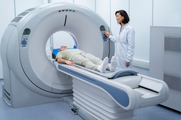
The CT scanner looks like a big ring or doughnut. You lie on a table, and it moves you through the ring while X-rays spin around your body.
A computer puts all those X-ray pictures together into cross-sectional (slice-like) images. This gives doctors a 3D view of the area.
That’s why people call CT a 3D X-ray. It shows much more detail than a normal X-ray.
B. How MRI Uses Magnets and Radio Waves
MRI stands for magnetic resonance imaging. The MRI machine is a large tube with a strong magnet inside.
When you lie inside, the magnet lines up the water molecules in your body. Then, radio waves shake them up for a second.
As the molecules relax, they send out signals. The scanner catches these and creates super-detailed images, especially of soft tissues.
MRI doesn’t use any X-rays.
Why CT Is Sometimes Called a CAT Scan? CAT scan means “computerized axial tomography.” “Axial” just describes the cross-sectional slices. People now say CT scan more often, but both mean the same thing.
The Biggest Differences That Matter to Patients
There’s more to it than just the technology. For you, the main differences are:
- How long the scan takes
- Whether it uses radiation
- Which body parts each scan shows best
A. Speed and Availability
CT scans are really fast. The scan itself often takes less than a minute.
Your whole appointment, including prep, might take 10–15 minutes.
MRI takes longer—usually 20–50 minutes, depending on the body part.
CT machines are found in most emergency rooms. That’s why doctors pick CT when time is critical.
B. Radiation Exposure and Safety
CT scans use ionizing radiation (like X-rays). Doctors say the risk from one scan is low, but they avoid it during pregnancy unless needed.
MRI scans don’t use any radiation. They’re safe for most people, even pregnant women in many cases.
Worried about radiation? Ask your doctor if MRI can answer the same question.
Image Detailings in Soft Tissue vs. Bone
Feature | CT Scan | MRI |
Best for | Bones, bleeding, organs, lungs | Brain, spine, joints, nerves |
Radiation | Yes (X-rays) | No |
Scan time | Under 1 minute | 20–50 minutes |
Noise level | Quiet | Loud clicking sounds |
Metal implants | Usually safe | Not always safe |
CT is great for dense things like bones and quick bleeding checks. MRI gives much better detail for soft tissues.
When a CT Scan Is Usually the Better Choice
CT is best when speed matters, or when doctors want to check bones, stones, bleeding, or organs. A radiologist can see the results within minutes.
A. Trauma, Internal Bleeding, and Emergencies
In the ER, doctors almost always use CT first. If you had a car accident, a bad fall, or signs of internal bleeding, CT finds the problem fast.
It detects bleeding in the brain, chest, or abdomen quickly. In emergencies, speed is everything.
B. Bone Injuries, Fractures, and Spine Evaluation
CT is excellent for seeing fractures, especially tricky ones. Surgeons use CT to get a 3D look before surgery.
For spinal injuries, CT shows the bones and any damage pressing on the spinal cord.
C. Kidney Stones, Lungs, and Fast Organ Imaging
Doctors call CT the expert at “bones and stones.” It’s the best way to spot kidney stones and gallstones, and shows their size and location.
CT is also used for:
- Checking lung problems and chest infections
- Staging cancer and seeing how far it’s spread
- Quickly checking abdominal organs in emergencies
- Planning for surgery
When an MRI Is Usually the Better Choice
MRI is best for soft tissues, the brain, spinal cord, nerves, or joints. Since MRI doesn’t use radiation, it’s safer if you need repeat scans.
A. Brain, Spine, and Nerve Conditions
MRI is the top choice for brain and spinal cord scans. If your doctor thinks you have a stroke, multiple sclerosis, epilepsy, a brain tumour, or a spine disc problem, MRI is almost always ordered.
MRI can also show nerves in some areas—CT can’t do that.
B. Joints, Muscles, Ligaments, and Other Soft Tissues
Got knee pain, shoulder trouble, or a torn ligament? MRI is what doctors order. It shows muscles, tendons, cartilage, and ligaments in much more detail than CT.
Sports injuries and joint issues—MRI gives the clearest picture.
C. Functional MRI and Advanced MRI Uses
Functional MRI (fMRI) goes further. It measures brain activity by tracking blood flow—mainly for research or brain surgery planning.
Other advanced MRI uses:
- Mapping blood vessels without dye
- Detecting early prostate cancer
- Checking heart muscle function
- Spotting tiny brain changes before symptoms start
What to Expect Before, During, and After the Exam
Knowing what happens at each step can help you relax. CT and MRI feel different, so here’s what you can expect.
A. Preparation and Screening
Before both scans, you’ll need to remove metal things—jewellery, glasses, hairpins.
For MRI, the staff will ask more questions because of the strong magnet. They’ll check for:
- Metal implants, pacemakers, or defibrillators
- Surgical clips or staples
- Old injuries with metal fragments
- Cochlear implants or drug pumps
Some implants are safe for MRI, some are not. Always tell the truth in screening.
For CT, you might need to skip food or drink for a few hours if contrast dye is needed. Pregnant women should always inform the staff.
B. Contrast Agents and When They Are Used
Both scans sometimes use contrast agents (dye injected into a vein) to make some areas clearer.
- CT contrast: iodine-based, highlights blood vessels, organs, tumours.
- MRI contrast: gadolinium-based, helps spot inflammation, tumours, odd tissues.
Not every scan needs contrast. Your doctor will decide. If you’ve had allergic reactions to dye or have kidney problems, tell the team.
C. Comfort, Noise, and Staying Still
CT scans are fast and quiet. You just lie on a table that moves through the ring. The scan takes less than a minute.
MRI is louder and takes longer. The machine makes knocking and clicking sounds. You’ll get earplugs or headphones.
You have to stay still for 20–50 minutes inside a narrow tube. If you’re claustrophobic, tell your doctor. Some places have open MRI machines, which are less cramped.
How Doctors Decide Which Test You Need
Doctors don’t just pick the “better” scan. They choose based on your symptoms, the body part, and what they need to find out. A radiologist and your doctor work together to pick the right scan.
A. Body Part and Symptoms Being Investigated
The main factor is where your symptoms are.
- Head injury or sudden bad headache: CT first for bleeding, then maybe MRI for more detail.
- Knee or shoulder pain: MRI is the usual first choice.
- Chest pain or breathing trouble: CT is often used.
- Suspected kidney stones: CT is most reliable.
- Possible nerve problem like MS or epilepsy: MRI is standard.
B. Questions to Ask Your Care Team
You should always know why you’re getting a scan. Try asking:
- Why this scan and not the other?
- Will you use contrast dye? Why?
- Any risks for me?
- Will you compare this scan to old ones?
- When will I get the results?
At a hospital like IPIMS, the radiology team can explain everything so you feel confident.
Frequently Asked Questions
Q. Which imaging test is better for my symptoms?
Neither scan is always better. It depends on your symptoms and what your doctor is looking for. CT is best for bones, bleeding, and emergencies. MRI is better for brain, spine, joints, and soft tissues.
Q. What are the main differences in how these scans work?
CT uses X-rays from many angles to make cross-sectional images. MRI uses a strong magnet and radio waves to make detailed images of soft tissues. No radiation is involved.
Q. How do doctors decide for head injuries or stroke symptoms?
For head injuries and stroke, CT comes first. It’s fast and shows bleeding. If more detail is needed, doctors order an MRI after that.
Q. Is one of these scans safer for radiation?
MRI doesn’t use radiation and is safe for most people, including many pregnant women. CT uses radiation, but one scan is usually considered low risk. Your doctor will balance the benefits and risks.
Q. How long does each scan take? What should I expect?
CT is very quick—scan done in under a minute, appointment over in 10–15 minutes. MRI takes longer—20–50 minutes, and you need to lie still inside a noisy, tube-like machine.
Still confused? Don’t worry. Ask your doctor or radiologist—they’re there to help you understand and make the right choice for your health.
Q. Can I have one of these scans if I have metal implants, braces, or a pacemaker?
- CT scans are usually safe for people with metal implants or braces.
- You can go for a CT even if you have most medical devices.
- MRI is a bit tricky. This scan uses a strong magnet.
- If you have a pacemaker, defibrillator, or cochlear implant, MRI might not be safe.
Always tell the technician if you have any metal in your body.
This is super important, especially before an MRI.