Orthopaedic Surgery including Joint
At IPIMS Hospital’s Orthopaedic Department in Panipat, we understand that bone, joint, and muscle pain doesn’t just affect your body it affects every part of your daily life. Whether you are struggling to climb stairs, suffering from a sports injury, or recovering from a fracture, our expert orthopaedic team is here to get you moving again.
Located conveniently on Main G.T. Road, Panipat, Haryana, IPIMS is one of the most trusted orthopaedic care centres serving patients from across Panipat, Karnal, Samalkha, Israna, and Kaithal.
Our department combines advanced surgical technology with compassionate, personalized care. From children with bone deformities to elderly patients needing joint replacements, we provide complete orthopaedic treatment under one roof no need to travel to Delhi or Chandigarh.
What Is Orthopaedic Care?
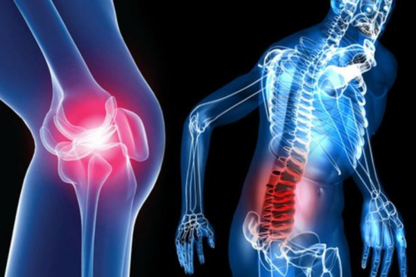
Elderly patients commonly suffer from arthritis, osteoporosis, and joint deterioration that makes walking painful. In Haryana, musculoskeletal disorders are among the leading causes of disability and reduced quality of life. Many patients in Panipat suffer silently for years, unaware that effective surgical and non-surgical treatments are available locally. At IPIMS, our orthopaedic specialists bring tertiary-level expertise to your doorstep, right here in Panipat.
How We Treat It:
Treatment depends entirely on the type and location of the fracture. Simple, stable fractures are managed with closed reduction the doctor manually realigns the bone without surgery followed by plaster casting or a splint to hold it in place while healing occurs over 4–8 weeks.
For more serious fractures such as compound (open) fractures or displaced bones Open Reduction and Internal Fixation (ORIF) surgery is performed. Our surgeons use metal plates, screws, or intramedullary nails inserted inside the bone to hold fragments in precise alignment. This is essential for fractures of the femur (thigh bone), tibia (shin bone), and wrist.
Complex poly-trauma cases involving multiple fractures common in road accidents are stabilized with external fixators first, then reconstructed in staged surgeries. Post-fracture physiotherapy at our in-house rehab unit ensures full restoration of strength and movement.
How We Treat It:
Treatment follows a step-by-step ladder from least to most invasive, based on severity.
In early-stage arthritis, we use a combination of anti-inflammatory medications, physiotherapy to strengthen the muscles supporting the knee, weight management counselling, and corticosteroid or hyaluronic acid (viscosupplementation) injections directly into the joint for pain relief and lubrication.
For patients not responding to conservative care, Platelet-Rich Plasma (PRP) therapy uses the patient’s own blood to stimulate cartilage healing and reduce inflammation.
In severe arthritis where the joint space is completely destroyed, Total Knee Replacement (TKR) surgery is the definitive solution. Our surgeons replace the damaged joint surfaces with precisely fitted metal and high-grade plastic components. Most patients begin walking with support within 24–48 hours after surgery, and full recovery takes 6–8 weeks with guided physiotherapy.
How We Treat It:
Mild to moderate hip pain from early arthritis or bursitis is managed with physiotherapy, anti-inflammatory medications, and steroid injections into the hip joint.
Avascular necrosis in early stages can be addressed with a procedure called core decompression — drilling tiny channels into the femoral head to restore blood supply and prevent further collapse of the bone.
For advanced hip arthritis or fractures in elderly patients, Total Hip Replacement (THR) is the gold-standard treatment. The damaged femoral head and acetabular socket are replaced with a metal stem, ceramic or metal ball, and a plastic or ceramic cup. This completely eliminates the source of pain. Patients are mobilized within 1–2 days post-surgery. Our implants are sourced from internationally certified manufacturers, ensuring longevity of 15–25 years.
How We Treat It:
The majority of back problems — nearly 85–90% — are successfully treated without surgery. Our first-line approach combines structured physiotherapy, hot/cold therapy, postural correction training, and NSAIDs or muscle relaxants to relieve acute pain. For persistent nerve pain from disc herniation, epidural steroid injections or nerve root block injections deliver anti-inflammatory medication directly to the compressed nerve, providing significant relief.
Surgical intervention is reserved for cases where nerve compression is causing weakness, numbness, or loss of bladder/bowel control. We perform Microdiscectomy (minimally invasive disc fragment removal), Laminectomy (spinal canal decompression), and Spinal Fusion using titanium implants for unstable vertebrae. Our spine surgeries are performed under advanced imaging guidance to ensure precision and safety.
How We Treat It:
Frozen shoulder is treated through a progressive physiotherapy programme combined with intra-articular corticosteroid injections. A procedure called Manipulation Under Anaesthesia (MUA) — where the shoulder is gently mobilized while the patient is sedated — can rapidly break adhesions and restore movement.
Partial rotator cuff tears are managed with physiotherapy and PRP injections to promote tendon healing. Complete rotator cuff tears in active patients are repaired using arthroscopic (keyhole) surgery — the torn tendon ends are sutured back to the bone using tiny anchors through two or three small incisions, avoiding large open cuts.
Recurrent shoulder dislocations are corrected with Bankart repair surgery (arthroscopic stabilization), tightening the ligaments that hold the shoulder joint in place. Recovery involves a sling for 4 weeks followed by progressive physiotherapy.
How We Treat It:
ACL (Anterior Cruciate Ligament) tears — the most common serious sports injury — are reconstructed arthroscopically. A graft (usually taken from the patient’s own hamstring or patellar tendon) replaces the torn ligament and is fixed inside the knee through tiny incisions. This is keyhole surgery — no large cuts, minimal blood loss, and the patient is typically discharged within 2 days.
Meniscus tears (cartilage damage inside the knee) are treated with arthroscopic meniscus repair or partial meniscectomy, depending on tear location and pattern. Tendon ruptures such as Achilles tendon tears are repaired surgically with suture techniques, followed by a structured rehabilitation protocol.
Our in-house physiotherapy unit runs sport-specific rehabilitation programmes to return athletes to competitive activity safely, reducing re-injury risk.
How We Treat It:
The first step is diagnosis using a Bone Mineral Density (BMD/DEXA) scan available at IPIMS, which measures bone density and predicts fracture risk. Treatment includes calcium and Vitamin D supplementation, dietary counselling, weight-bearing exercise programmes, and fall prevention strategies at home.
For patients with confirmed osteoporosis, anti-resorptive medications (bisphosphonates such as alendronate or zoledronic acid infusions) are prescribed to slow bone loss and strengthen existing bone. Anabolic agents like teriparatide are used in severe cases to actively build new bone tissue.
If a vertebral fracture has already occurred causing severe back pain, a minimally invasive procedure called Vertebroplasty or Kyphoplasty — injecting bone cement into the collapsed vertebra — provides immediate pain relief and restores vertebral height.
How We Treat It:
Club foot in newborns is treated with the internationally proven Ponseti Method — a series of weekly plaster casts progressively correct the foot position over 6–8 weeks, often avoiding surgery. A minor Achilles tendon release procedure and a foot abduction brace complete the treatment.
Developmental dysplasia of the hip (DDH) in infants is treated with a Pavlik harness — a soft brace worn for 6–8 weeks — to guide the hip into correct alignment. Older children may require surgery.
Limb length discrepancies are corrected using guided growth techniques (temporary stapling) or limb lengthening with an Ilizarov / Taylor Spatial Frame for larger discrepancies.
Scoliosis is monitored with serial X-rays; mild curves are managed with bracing, while progressive curves above 40–45° require surgical correction with spinal rods.
How We Treat It:
Mild to moderate cases are managed non-surgically. A wrist splint worn at night keeps the wrist in a neutral position, relieving pressure on the nerve. Corticosteroid injections into the carpal tunnel reduce inflammation and provide significant short-term relief for most patients.
When symptoms are severe, long-standing, or not responding to conservative treatment, Carpal Tunnel Release surgery is performed. This is a simple, day-care (outpatient) procedure done under local anaesthesia — the transverse carpal ligament is divided, immediately releasing the compressed nerve.
The procedure takes under 30 minutes. Patients typically notice improvement in tingling and numbness within days, with full recovery in 3–4 weeks.
How We Treat It:
Acute osteomyelitis (recent infection) is treated aggressively with high-dose intravenous antibiotics targeting the causative organism — identified through blood cultures and bone biopsy. Antibiotic treatment typically continues for 4–6 weeks, transitioning to oral antibiotics for a further 4–8 weeks.
When infection has caused bone destruction, dead bone tissue (called a sequestrum) must be surgically removed in a procedure called sequestrectomy and debridement. The cavity left behind may be filled with antibiotic-impregnated calcium sulphate beads that release antibiotics locally over weeks, ensuring sterilization of the infected site without systemic side effects.
In diabetic foot osteomyelitis, treatment is multidisciplinary — our orthopaedic team works closely with diabetologists and vascular surgeons to achieve infection control and limb salvage, avoiding unnecessary amputation.
How We Treat It:
Mild-to-moderate sprains (Grade I & II) are treated with the PRICE protocol — Protection, Rest, Ice, Compression, and Elevation — combined with physiotherapy to restore strength and proprioception (joint sense). NSAIDs reduce inflammation and pain in the acute phase.
PRP (Platelet-Rich Plasma) injections into chronically injured tendons accelerate healing by delivering a high concentration of growth factors directly to damaged tissue, particularly useful for chronic Achilles tendinopathy or patellar tendinopathy.
Complete tendon ruptures (Grade III) — such as a total Achilles tendon tear — require surgical repair. The torn tendon ends are sutured together through a minimally invasive incision, followed by casting and a graduated rehabilitation programme. Return to full activity typically takes 4–6 months.
Chronic ankle instability from repeated sprains is corrected with ligament reconstruction surgery (Brostrom procedure), tightening the lateral ankle ligaments to prevent further episodes.
How We Treat It:
Evaluation begins with X-ray, MRI, and CT scanning to assess tumour size, location, and involvement of surrounding tissues. A bone biopsy — taking a small tissue sample — confirms the exact tumour type and guides treatment.
Benign tumours (e.g., osteochondromas, simple bone cysts) that are not causing symptoms may simply be monitored with periodic imaging. Symptomatic benign tumours are treated with surgical excision and bone grafting to fill the defect. Malignant bone tumours require a multidisciplinary approach. Our orthopaedic oncology team coordinates with medical oncologists and radiation oncologists.
Treatment typically combines neo-adjuvant chemotherapy (to shrink the tumour before surgery), followed by limb-salvage surgery (removing the tumour while preserving the limb using bone grafts or custom metal implants), and adjuvant chemotherapy or radiotherapy post-surgery. Our goal is always to preserve the limb and function wherever oncologically safe to do so.
Our Orthopaedic Services in Panipat
A. Diagnostic Services
- X-Ray (Digital): First-line imaging for fractures, arthritis, and bone alignment
- MRI Scan: Detailed imaging of soft tissues, ligaments, cartilage, and spinal discs; essential for diagnosing ACL tears, disc herniations, and early arthritis
- CT Scan: 3D bone imaging for complex fractures and pre-surgical planning
- Bone Mineral Density (BMD / DEXA Scan): Diagnoses osteoporosis and fracture risk, especially important for women above 45 in Haryana
- Ultrasound: Assessment of tendons, soft tissue swellings, and guided injections
- Laboratory Tests: Uric acid, CRP, ESR, RA factor, vitamin D, and calcium levels for arthritis and metabolic bone disease workup
- Nerve Conduction Study (NCS): For diagnosing carpal tunnel syndrome and nerve compression conditions
B. Surgical Treatments & Procedures
Joint Replacement Surgery
- Total Knee Replacement (TKR) — for severe knee arthritis
- Total Hip Replacement (THR) — for hip arthritis and femoral neck fractures
- Partial Knee Replacement (Unicompartmental) — when only part of the knee is affected
- Shoulder Replacement — for complex shoulder arthritis
Arthroscopic (Keyhole) Surgery
- ACL Reconstruction — for torn anterior cruciate ligament
- Meniscus Repair and Meniscectomy — for cartilage tears
- Shoulder Arthroscopy — rotator cuff repair, Bankart repair for instability
- Knee Arthroscopy — cartilage smoothing, loose body removal
Fracture Management
- Closed Reduction and Casting — for stable fractures
- Open Reduction and Internal Fixation (ORIF) — using plates, screws, and nails
- External Fixation — for complex, open, or infected fractures
- Intramedullary Nailing — for long bone fractures (femur, tibia)
Spine Surgery
- Microdiscectomy — minimally invasive removal of herniated disc material
- Spinal Fusion — for unstable vertebrae and severe spondylolisthesis
- Laminectomy — decompression of spinal canal in stenosis
Non-Surgical Treatments
- Physiotherapy and rehabilitation (in-hospital physio unit)
- Platelet-Rich Plasma (PRP) injections — for cartilage repair and sports injuries
- Corticosteroid joint injections — for rapid pain relief in arthritis
- Custom orthotics and bracing
- Traction therapy for spine
Why Patients in Panipat Trust IPIMS for Orthopaedic Care
Panipat patients no longer need to spend time, money, and energy travelling to Delhi or Chandigarh for quality orthopaedic treatment. Here is why thousands of patients from across Haryana choose IPIMS:
- Advanced Operation Theatres — Modern modular OTs equipped for joint replacement, arthroscopy, and trauma surgery with infection control standards
- In-House Imaging — MRI, CT Scan, and Digital X-Ray available on campus, reducing diagnosis delays
- 24/7 Orthopaedic Emergency — Road accident trauma, fractures, and acute joint injuries handled around the clock by trained staff
- Physiotherapy & Rehabilitation Unit — In-hospital physio ensures patients recover faster without needing to go elsewhere
- Affordable & Transparent Pricing — We offer clear, upfront treatment costs with no hidden charges; cashless facility for major insurance companies and Ayushman Bharat
- Central GT Road Location — Easily accessible from Panipat city, Samalkha, Israna, Madlauda, and Karnal — no long travel required
- Bilingual Care — Our doctors and staff communicate in Hindi and Haryanvi, so patients always understand their diagnosis and treatment plan
- Patient-First Philosophy — From your first consultation to post-operative recovery, our team walks with you every step of the way
Your Orthopaedic Treatment Journey at IPIMS
We know that visiting a hospital can feel overwhelming — especially when you're in pain. Here's exactly what to expect when you come to IPIMS for orthopaedic care:
Book Your Appointment
Call our helpline, book online through the IPIMS website, or walk into our OPD on Main GT Road, Panipat. Our reception team will guide you to the Orthopaedic OPD with zero waiting confusion.
Initial Consultation
Your orthopaedic specialist will take a detailed history, examine the affected area, and understand your lifestyle, occupation, and activity level — because treatment is always personalized. Bring any previous X-rays, reports, or prescriptions.
Diagnostics
If needed, tests like X-ray, MRI, blood work, or bone density scan are ordered. Since all diagnostic services are in-house at IPIMS, most reports are ready the same day or within 24 hours.
Diagnosis & Treatment Plan
Your doctor will explain your condition in simple language (Hindi or English), show you your imaging reports, and present all treatment options — from physiotherapy to surgery. You will never be pushed into a procedure without full understanding and consent.
Treatment / Surgery
If surgery is required, our team handles all pre-operative preparation, anaesthesia counselling, and surgery. Our modular OTs and trained nursing staff ensure safety and sterility at every step.
Recovery, Physiotherapy & Follow-Up
Post-surgery, our in-hospital physiotherapy team begins rehabilitation — helping you regain strength and movement. You will receive a clear discharge plan with follow-up dates, home exercise instructions, and direct doctor access for any concerns.
When Should You See an Orthopaedic Doctor in Panipat?
Many patients delay orthopaedic care, hoping pain will go away on its own. This often leads to conditions worsening and requiring more complex treatment. Watch for these warning signs:
- Persistent joint pain lasting more than 2 weeks
- Morning stiffness in knees, hips, or hands that takes more than 30 minutes to resolve
- Swelling in any joint without a clear injury
- Difficulty walking, climbing stairs, or standing from a seated position
- Numbness or tingling in arms or legs
- Back pain that radiates down the leg (possible sciatica)
- Bones or joints that crack, lock, or “give way”
- Reduced range of motion in any joint
- A bone fracture or suspected fracture after a fall or accident
- Severe sudden joint pain with inability to move a limb
- Open wound with visible bone or joint exposure
- Joint dislocation (shoulder popped out, kneecap displaced)
- Loss of sensation or movement in a limb after a spine injury
Orthopaedic Emergency at IPIMS Panipat — 24/7
📞 Call: +91 70566 67979
Our trauma and orthopaedic team is available around the clock.Frequently Asked Questions Orthopaedic Department at IPIMS Panipat
Visit IPIMS Orthopaedic Department — Panipat
Getting expert orthopaedic care in Panipat has never been easier. IPIMS Hospital is centrally located on Main G.T. Road, Panipat, Haryana, making it easily reachable whether you are coming from Panipat City, Samalkha, Israna, Madlauda, or from nearby Karnal and Sonipat.
📍 Address
IPIMS Hospital, Main G.T. Road, Panipat, Haryana
🕘 OPD Hours
Monday–Saturday, 09:00 AM – 05:00 PM
🚨 Emergency & Trauma
24 Hours, 7 Days a Week
📞 Phone
+91 70566 67979
🌐 Website
www.ipimshospital.com
📅 Book Your Orthopaedic Appointment Today
Don't let joint pain, back pain, or an untreated injury hold you back from living your full life. Our orthopaedic specialists in Panipat are ready to help you get back on your feet.